The cornea
The cornea is the clear ”windshield“ of the eye and as one of the refractive media it is significantly involved in the projection of the perceived objects.
Incident light passes through the tear film, is refracted by the cornea and continues through the aqueous fluid of the anterior chamber and through the pupil. It is subsequently refracted by the lens and passes through the vitreous to meet the retina. At this level light is processed into nerve impulses and transmitted to the visual center of the brain.
The cornea of the eye is the only transparent tissue of the human body and can therefore be seen as a miracle of nature. It is approximately 0.5 mm thick (in the center) and consists of 5 layers or membranes.
Keratoconus and its related forms
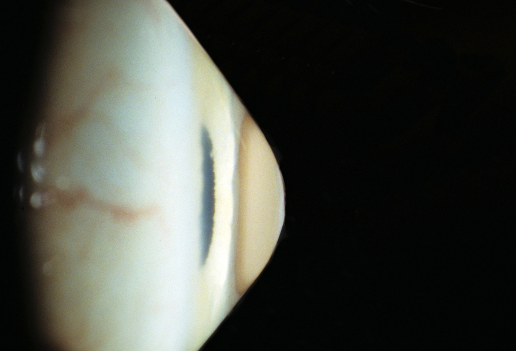
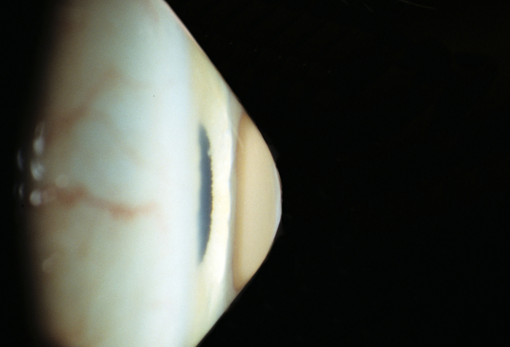
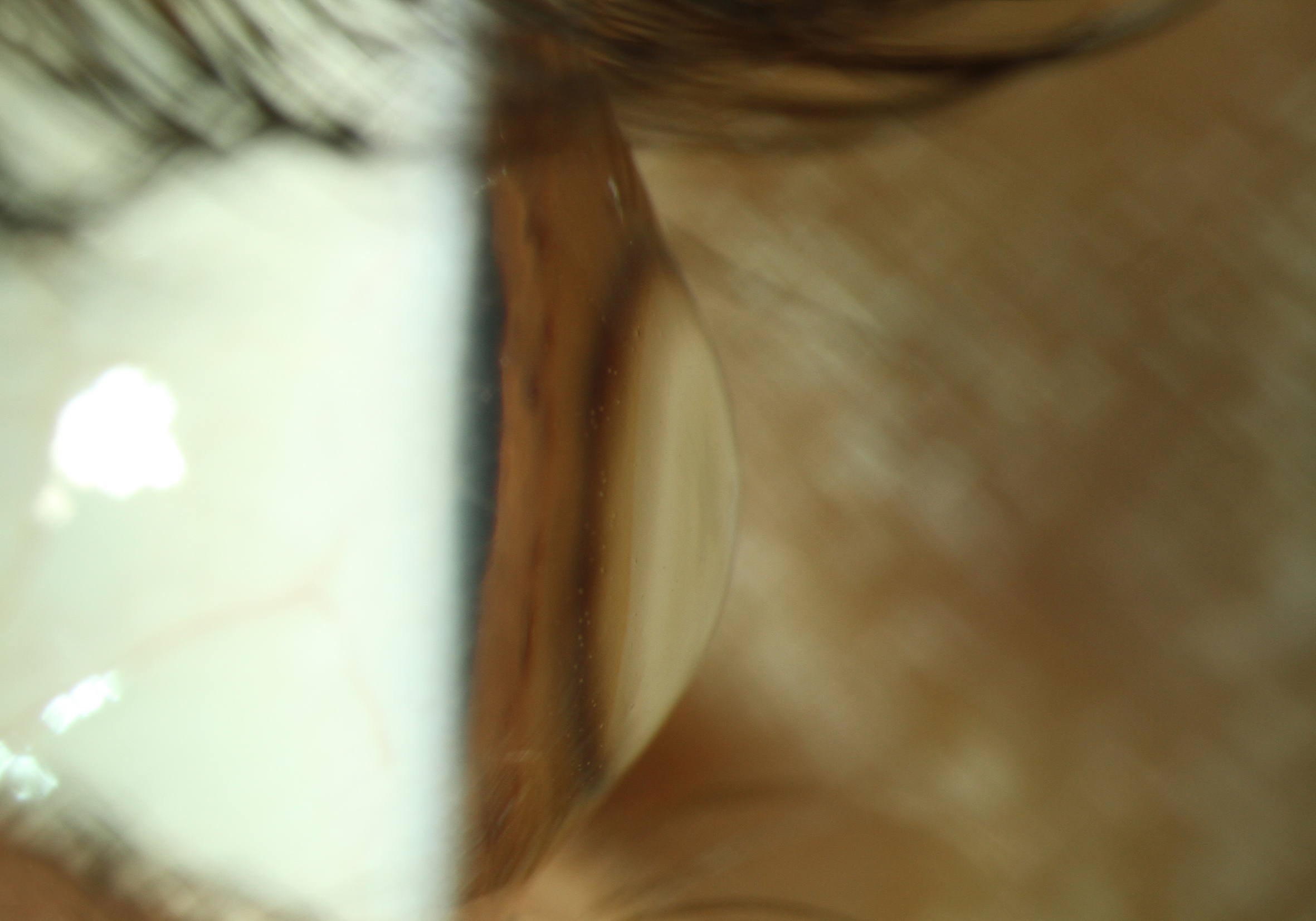
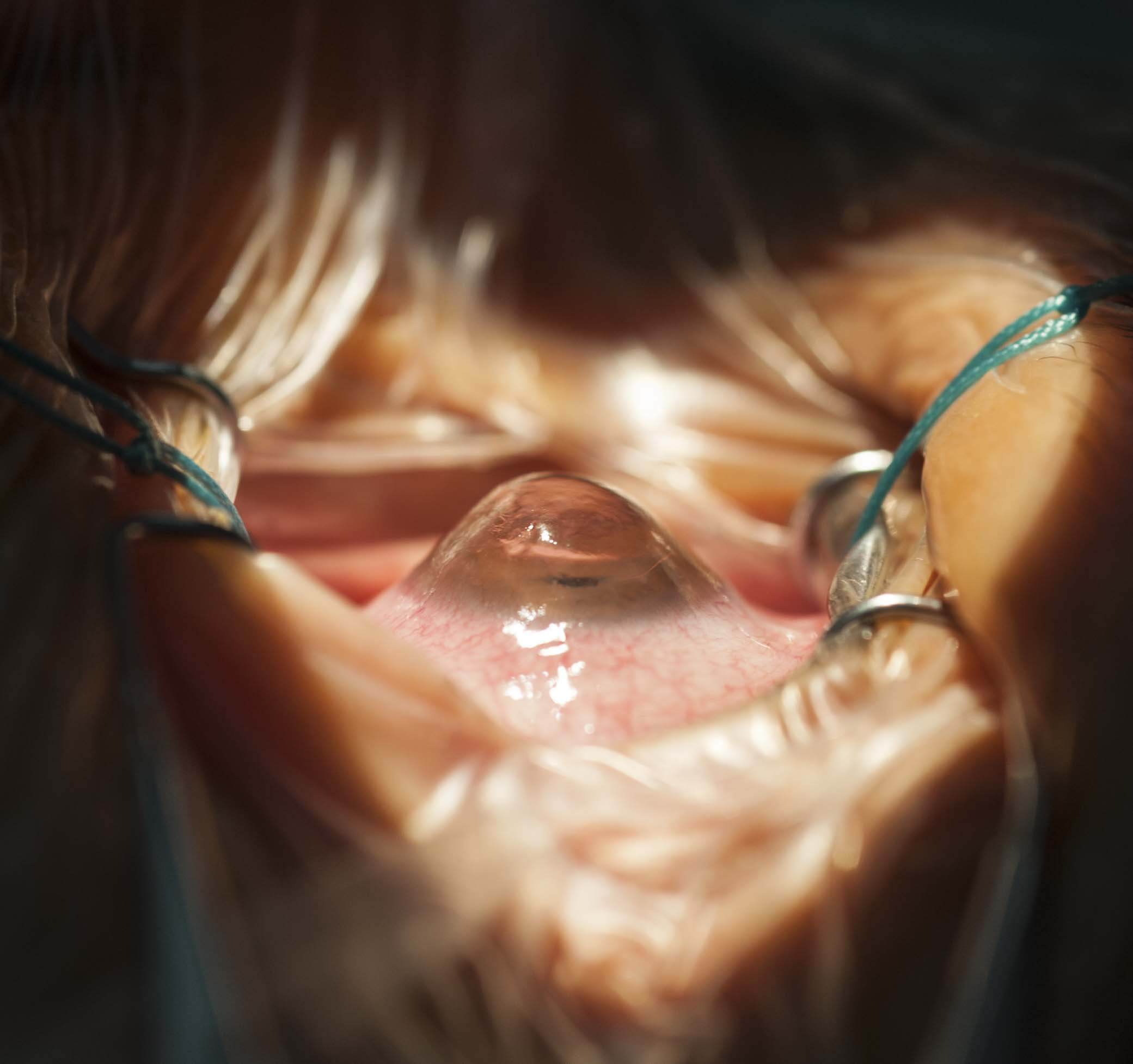
The term is composed of “keras” (Greek for horn) and “konus” (Latin for cone) and refers to a conical curvature of the cornea.
Changes which cause a deformation of the clear cornea of the eye are called ectatic diseases. They are generally progressive, however, the individual rate of progression is variable.
Characteristically, only the center of the cornea becomes weakened in keratoconus. In contrast, a generalized thinning with globular protrusion is called keratoglobus. An extreme band of thinning of the inferior cornea is referred to as keratotorus or pellucid marginal degeneration.
Keratoconus and Vision
Due to the increasing irregularity of the front and back corneal surface the quality of vision becomes impaired.
Patients often complain of blurry vision, increased sensitivity to light and poor night vision.
In most cases the vision can initially be optimized with corrective lenses. In advanced stages spectacles may not provide a satisfactory level of visual acuity while rigid gas-permeable contact lenses can achieve a good visual correction. Rigid gas-permeable contact lenses create a “tear lens” by means of tear fluid filling the gap between the back of the lens and the irregular corneal surface and thus improve visual acuity and reach an almost “normal” vision.
Clinical Diagnostics
Ectatic corneal diseases are rather common with 1 in 2000 people being affected in Europe and the USA. They are hardly recognizable and therefore often not diagnosed in the early stages.
A frequently changing prescription of glasses over a short period of time should raise suspicion for ectatic diseases.
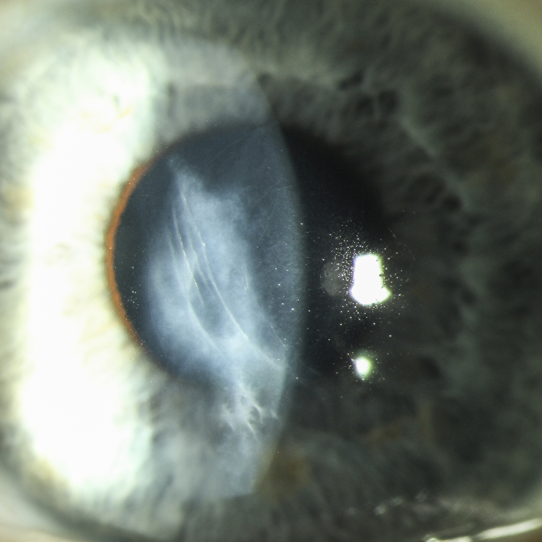
The early stage of keratoconus cannot reliably be diagnosed by means of slit lamp examination of the cornea. A computer-based 2-dimensional curvature image called “corneal topographic analysis” is required.
In addition, the analysis of corneal rigidity by means of the Ocular Response Analyzer (ORA) by using the Keratoconus Match Index (KMI) or by means of CORVIS can be helpful in case of doubt or for further differentiation.
Treatment of Keratoconus
Currently, there is no cure for keratoconus. The thinning and protrusion of the cornea result from a biomechanical weakness.
Today, microsurgical interventions are able to slow disease progression. These procedures include corneal collagen cross-linking (Riboflavin-UVA-Crosslinking) which stiffens the cornea to prevent further shape changes and stop disease progression.
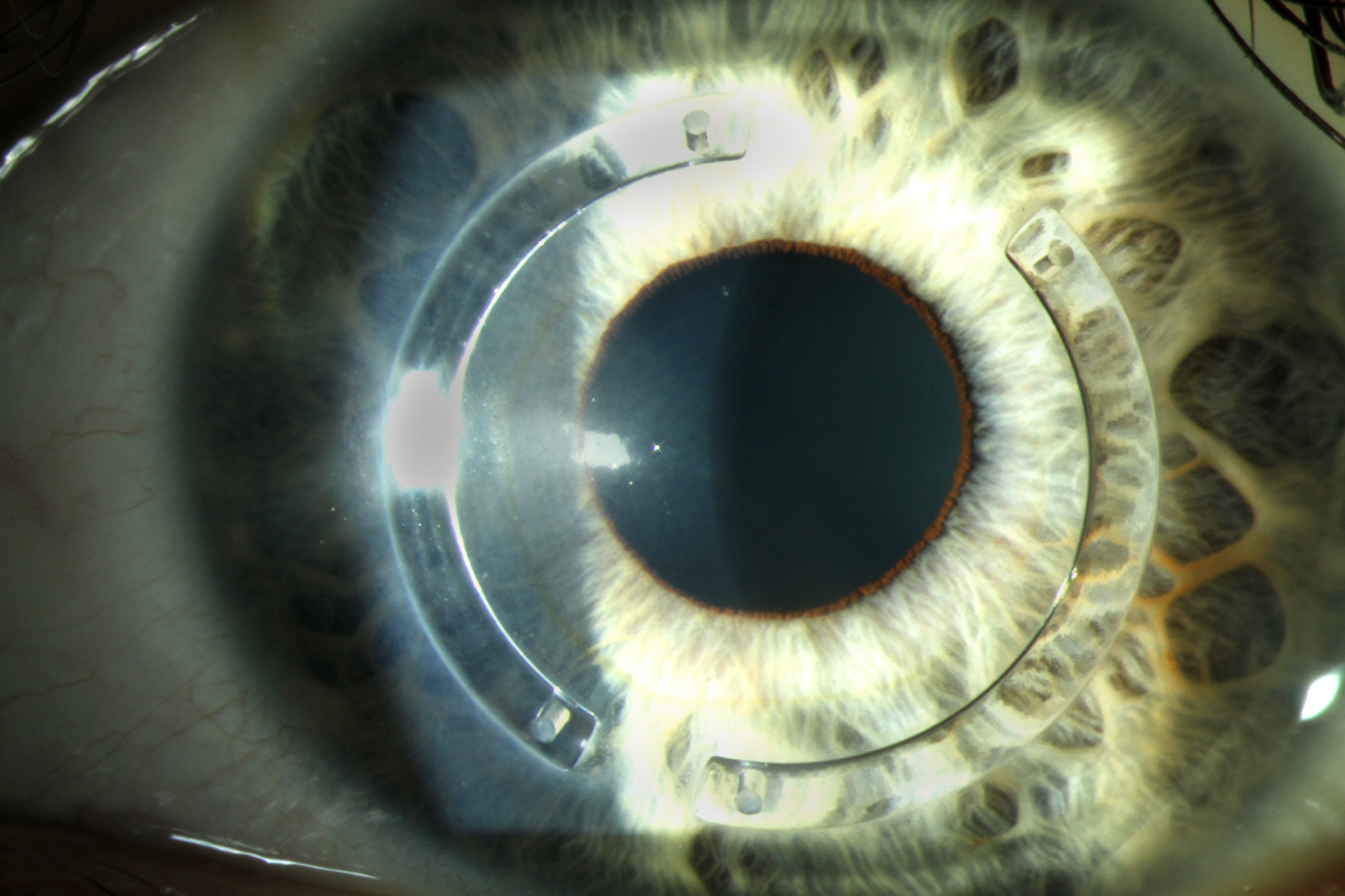
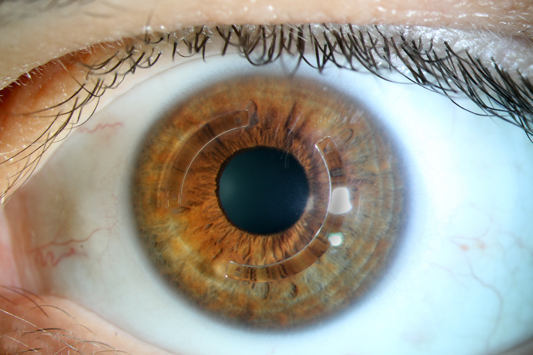
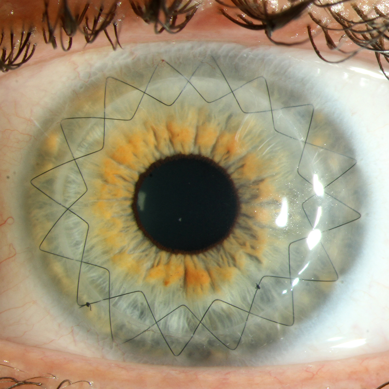
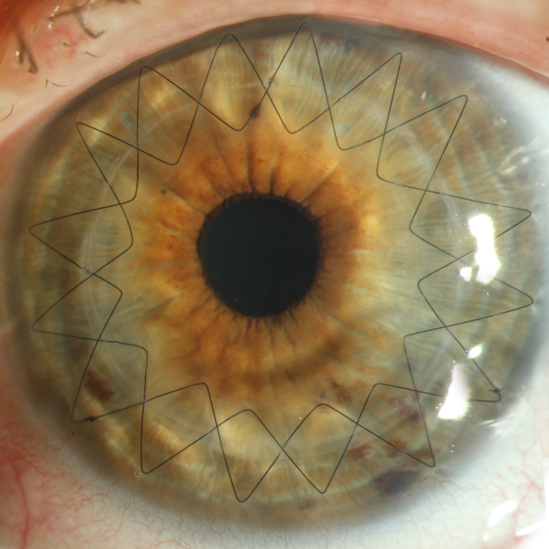
The implantation of nearly invisible ring segments made of synthetic material into the peripheral cornea is another option. They cause a flattening and regularization of the corneal surface and may stabilize disease progression. In advanced cases, lamellar or penetrating excimer laser keratoplasty (=corneal transplantation) is the only option. The thinned and scarred tissue is replaced by a clear donor tissue.
Research
Scientists and physicians have not yet been able to find the reason for keratoconus and its related forms. It appears to be genetic in some cases but exogenous factors like excessive eye rubbing seem to be relevant as well. Keratoconus is often associated with diseases like neurodermitis, allergies, Down`s syndrome (Trisomy 21) and hypothyroidism.
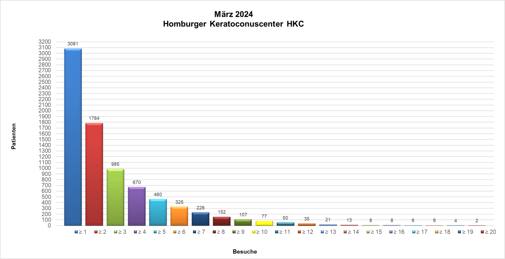
Here you will find Publications (Pubmed) about the Homburg Keratoconus Center HKC.
- 2021. Hypoxic stress increases NF-κB and iNOS mRNA expression in normal, but not in keratoconus corneal fibroblasts
- hypoxic-stress-increases-nf-b-and-inos-mrna-expression-in-normal-but-not-in-keratoconus-corneal-fibroblasts.pdf [800.23 KB]
- 2021. NF-κB, iNOS, IL-6, and collagen 1 and 5 expression in healthy and keratoconus corneal fibroblasts after 0.1% riboflavin UV-A illumination
- nf-b-inos-il-6-and-collagen-1-and-5-expression-in-healthy-and-keratoconus-corneal-fibroblasts-after-0.1-riboflavin-uv-a-illumina.pdf [1.44 MB]
- 2021. Tomographically normal partner eye in very asymmetrical corneal ectasia: biomechanical analysis
- tomographically-normal-partner-eye-in-very-asymmetrical-corneal-ectasia-biomechanical-analysis.pdf [420.69 KB]
- 2021. excimer laser-assisted DALK: A case report from the Homburg Keratoconus Center (HKC).
- excimerlaser-gestuetzte-dalk-ein-fallbericht-aus-dem-homburger-keratokonus-center-hkc.pdf [471.57 KB]
- 2020. Keratoconus staging by decades: a baseline ABCD classification of 1000 patients in the Homburg Keratoconus Center
- keratoconus-staging-by-decades-a-baseline-abcd-classification-of-1000-patients-in-the-homburg-keratoconus-center.pdf [618.72 KB]
- 2020. keratoconus detection and derivation of the degree of expression from the parameters of the Corvis®ST.
- keratokonusdetektion-und-ableitung-des-auspraegungsgrades-aus-den-parametern-des-corvis-st.pdf [651.82 KB]
- 2020. 63-year-old patient with acute visual deterioration after penetrating keratoplasty for keratoconus.
- ein-63-jaehriger-patient-mit-akuter-sehverschlechterung-nach-perforierender-keratoplastik-bei-keratokonus.pdf [746.79 KB]
- correlation-between-corneal-endothlial-cell-density-and-central-ocular-surface-temperature.pdf
- correlation-between-corneal-endothlial-cell-density-and-central-ocular-surface-temperature.pdf [433.45 KB]
- 2020. Ocular surface disease index and ocular thermography in keratoconus patients
- ocular-surface-disease-index-and-ocular-thermography-in-keratoconus-patients.pdf [1.1 MB]
- 2020. reliability of corneal tomography after implantation of intracorneal ring segments in keratoconus.
- reliabilitat-der-hornhauttomografie-nach-implantation-von-intrakornealen-ringsegmenten-bei-keratokonus.pdf [647.24 KB]
- 2020. Structural changes in the corneal subbasal nerve plexus in keratoconus
- structural-changes-in-the-corneal-subbasal-nerve-plexus-in-keratoconus.pdf [611.9 KB]
- 2019. Comparison of excimer laser versus femtosecond laser assisted trephination in penetrating keratoplasty
- comparison-of-excimer-laser-versus-femtosecond-laser-assisted-trephination-in-penetrating-keratoplasty-a-retrospective-study.pdf [333.89 KB]
- 2019. muraine sutures accelerate healing of hydrops corneae in acute keratotorus.
- muraine-nahte-beschleunigen-die-abheilung-des-hydrops-corneae-bei-akutem-keratotorus.-pdf.pdf [1.15 MB]
- 2019. Hypothyroidism is not associated with keratoconus disease
- hypothyroidism-is-not-associated-with-keratoconus-analysis-of-626-subjects.pdf [1.08 MB]
- 2018. Keratoconus-like tomographic changes in a case of recurrent interstitial keratitis
- keratoconus-like-tomographic-changes-in-a-case-of-recurrent-interstitial-keratitis.pdf [569.56 KB]
- 2018. Keratoconus associated with cornea guttata - Implication for disease progression
- keratoconus-associated-with-cornea-guttata-implications-for-disease-progression.pdf [137.15 KB]
- 2018. Endothelial alterations in 712 keratoconus patients
- endothelial-alterations-in-712-keratoconus-patients.pdf [228.97 KB]
- 2017. Penetrating keratoplasty for keratoconus excimer versus femtosecond laser trephination
- penetrating-keratoplasty-for-keratoconus-excimer-versus-femtosecond-laser-trephination.pdf [778.96 KB]
- 2017. Intraindividual Keratoconus Progression
- keratokonusprogression-im-seitenvergleich.pdf [89.55 KB]
- 2017. Complementary keratoconus indices based on topographical interpretation of biomechanical waveform parameters
- complementary-keratoconus-indices-based-on-topographical-interpretation-of-biomechanical-waveform-parameters-a-supplement-to-est.pdf [973.94 KB]
- 2016. Role of thyroxine in the development of keratoconus
- role-of-thyroxine-in-the-development-of-keratoconus.pdf [757.2 KB]
- PKP for Keratoconus – From Hand/Motor Trephine to Excimer Laser and Back to Femtosecond Laser
- perforierende-keratoplastik-bei-fortgeschrittenem-keratokonus-vom-hand-und-motortrepan-zum-excimerlaser-und-zuruck-zum-femtoseku.pdf [947.29 KB]
- 2016. Intracorneal Ring Segments to Treat Keratectasia – Interim Results and Potential Complications
- intrakorneale-ringsegmente-bei-keratektasien-zwischenergebnisse-und-potentielle-komplikationen.pdf [170.54 KB]
- 2016. stage-appropriate therapy of keratoconus and Oskar Fehr Lecture.
- stadiengerechte-therapie-des-keratokonus-editorial.pdf [142.98 KB]
- 2015. influence of individual microsurgical technique on endothelial cell density and pachymetry after penetrating keratoplasty in patients with Fuchs dystrophy or keratoconus.
- einfluss-der-individuellen-mikrochirurgischen-technik-auf-endothelzelldichte-und-pachymetrie-nach-perforierender-keratoplastik-b.pdf [109.81 KB]
- 2015. Excimer versus femtosecond laser assisted penetrating keratoplasty in keratoconus and fuchs dystrophy:
- excimer-versus-femtosecond-laser-assisted-penetrating-keratoplasty-in-keratoconus-and-fuchs-dystrophy-intraoperative-pitfalls.pdf [1.42 MB]
- A Comparison of Device-Based Diagnostic Methods for Keratoconus
- gerategestutzte-diagnostikverfahren-des-keratokonus-im-vergleich.pdf [450.64 KB]
- 2014. Diagnostic capacity of biomechanical indices from a dynamic bidirectional applanation device in pellucid marginal degeneration
- diagnostic-capacity-of-biomechanical-indices-from-a-dynamic-bidirectional-applanation-device-in-pellucid-marginal-degeneration.pdf [643.29 KB]
- 2014. Diagnostic capacity of the keratoconus match index and keratoconus match probability in subclinical keratoconus
- diagnostic-capacity-of-the-keratoconus-match-index-and-keratoconus-match-probability-in-subclinical-keratoconus.pdf [294.01 KB]
- 2013. DALK and penetrating laser keratoplasty for advanced keratoconus.
- dalk-und-perforierende-laserkeratoplastik-bei-fortgeschrittenem-keratokonus.pdf [895.91 KB]
- 2013. the little correction miracle
- keratokonuslinse-das-kleine-korrektionswunder.pdf [672.3 KB]
- 2013. news on the biomechanics of the cornea in keratoconus.
- neues-zur-biomechanik-der-kornea-bei-keratokonus.pdf [930.58 KB]
- 2013. intracorneal ring segments in keratoconus.
- intrakorneale-ringsegmente-beim-keratokonus.pdf [654.12 KB]
