Properties, extraction, conservation and delivery
The properties of AM on the cornea and conjunctiva include:
• stimulation of re-
epithelialization
• anti- inflammatory effects • anti-infectious effects
• immunomodulatory effects
• inhibition of corneal neovascularization
• inhibition of scar formation
• promotion of renervation
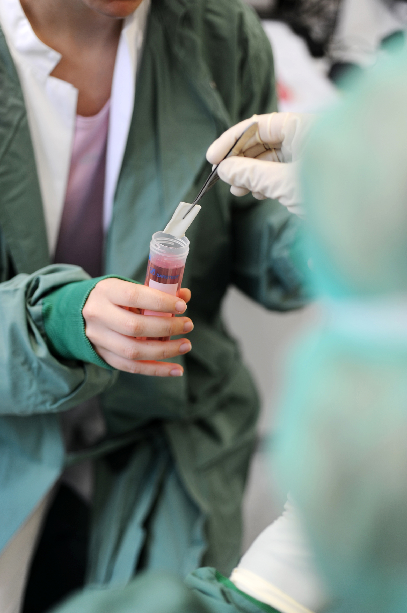
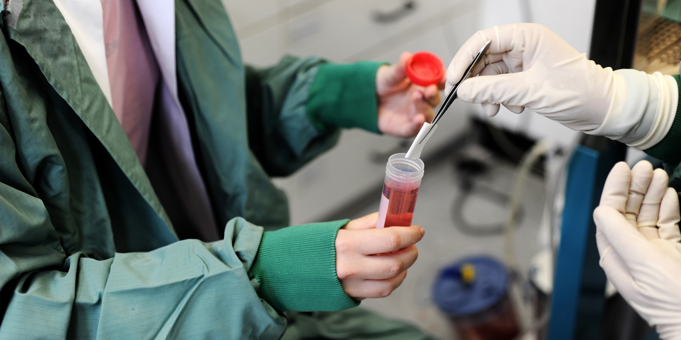
The acquisition of AM is exclusively after cesarean. After the donor has given consent to the tissue removal and the anamnestic clarification of the contraindications by a doctor, the tissue removal is carried out in close cooperation with the gynecology department at the UKS. The AM is applied to a carrier film after manual separation from the chorion. The tissue preservation is effected by kryokonservierende storage at temperatures between -75 ° C and -85 ° C.
Before a batch of human amniotic membrane, cryopreserved, Homburg/Saar is released, a comprehensive contraindication, serological and molecular biological testing of the donor and sterility testing of the media, rinsing solutions and the processed and unprocessed tissue takes place.
Transport and discharge of the amniotic membrane effected according to strict quality standards after a written agreement between the tissue bank and the clinical user / operator, and require a precise identification of the transport containers, and a transport of the cryopreserved AM frozen on dry ice (min. -79 ° C).
Each membrane is sent with instructions for use and specialist information.
Indications and application techniques
Typical indications for AM transplantation (AMT) are:
• persistent corneal epithelial defects
• epibulbar / tarsal conjunctival defects
• acute burns
• severe recurrent pterygium with formation of symblepharons
• before / after / simultaneously with keratoplasty
• ex vivo expansion of limbal stem cells as a carrier
Surgical techniques/possible uses of AMT for corneal defects:
• Patch (overlay): for epithelial defects or flat current defects:
The AM with a diameter of 16 mm is episclerally fixed in a circular manner (with continuous suture and contact lenses). The AM falls off after 1-2 weeks with no residuals, the contact lens and sutures are removed after 4 weeks.
• Graft (inlay,
single or multi-layer): The AM with a smaller diameter than the defect is sutured corneally into the stroma (top graft layer with several lamellar 10/0 nylon single sutures) and serves as a basement membrane over which the epithelium is formed grows. The AM is integrated into the cornea.
• Sandwich: combination of graft and patch technique.

Amnion harvesting at the UKS
An amniotic membrane for use on the eye was prepared for the first time in 2004 in the LIONS corneal bank Saar-Lor-Lux, Trier / West Palatinate. In 2010 the corneal bank was granted the so-called “manufacturing license” according to § 20b and § 20c of the Medicines Act by the Saarland Ministry of Health for both the amniotic membrane and the corneal transplants.
The corneal bank received permission from the Paul Ehrlich Institute in Langen in 2012 for corneas and in August 2015 for amniotic membranes in accordance with Section 21a of the German Medicines Act, which allows transplants to be given to other clinics and operating room centers.
